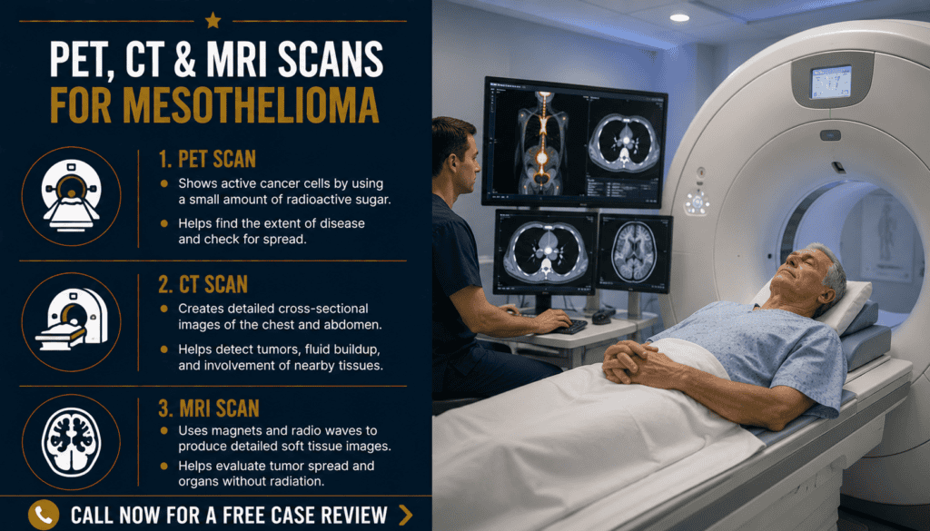
PET, CT & MRI Scans for Mesothelioma: What Each Test Shows
A practical patient guide to imaging tests, staging, and the questions to ask your care team.
Imaging helps doctors see where suspected or confirmed mesothelioma is located, estimate how far it extends, choose a safe biopsy location, and plan care. CT, PET/CT and MRI answer different questions. No scan alone reliably proves that a person has mesothelioma: tissue examination by a pathologist is generally needed for confirmation.
Why doctors order imaging
Mesothelioma most often affects the pleura, the tissue around the lungs, or the peritoneum, the lining of the abdomen. A scan may reveal pleural thickening, fluid, a mass, enlarged lymph nodes or other abnormalities. Similar findings can arise from other conditions, so imaging is interpreted together with symptoms, exposure history, examination and pathology.
Doctors use imaging at several points: investigating symptoms, selecting biopsy sites, estimating disease extent (staging), assessing treatment options, and comparing later scans with a baseline. The sequence depends on where the disease is suspected and the clinical question.
| Clinical question | How imaging may help | Important limitation |
|---|---|---|
| What is causing chest symptoms? | Chest CT can show pleural abnormalities and fluid. | Imaging cannot establish the cell type. |
| Has cancer spread? | CT and sometimes PET/CT assess regional and distant sites. | Suspicious sites may require tissue sampling. |
| Is a nearby structure invaded? | MRI may clarify chest-wall or diaphragm involvement. | Not routinely necessary for everyone. |
| Has treatment changed the disease? | Comparable follow-up CT images may help assess change. | Scarring and inflammation can complicate interpretation. |
PET vs. CT vs. MRI: the main differences
The tests complement one another rather than compete. A clinician chooses the smallest useful set of examinations for the patient’s situation. The following overview primarily concerns pleural mesothelioma; imaging plans for abdominal disease can differ.
| Scan | What it measures | Common role | Potential downside |
|---|---|---|---|
| CT | X-ray-based detailed anatomy | Usually the initial cross-sectional staging study of chest and upper abdomen, often with IV contrast | Radiation; iodinated contrast may be unsuitable for some patients |
| FDG PET/CT | Glucose-tracer activity combined with CT anatomy | May reveal metabolically active nodes or distant disease, particularly when staging changes treatment | Radiation; infection/inflammation can cause uptake |
| MRI | Magnetic-field images emphasizing soft tissues | Selected cases: define possible diaphragm, chest-wall or mediastinal invasion | Longer exam; metal implants and gadolinium require screening |
CT scans: the usual starting point
Computed tomography creates cross-sectional images of the chest and, when appropriate, upper abdomen. For pleural mesothelioma, contrast-enhanced CT is commonly used to map pleural thickening, fluid, adjacent structures and visible lymph nodes. CT may also help the team decide where a biopsy can be performed safely.
CT findings may include nodular or circumferential pleural thickening, a pleural effusion, or changes in lung volume. None is exclusive to mesothelioma. A normal or indeterminate scan does not settle every clinical question when symptoms or other findings remain concerning.
| What the report may mention | Plain-language meaning |
|---|---|
| Pleural thickening | The lining around the lung looks thicker than expected. |
| Pleural effusion | Fluid has collected between pleural layers. |
| Mediastinal lymphadenopathy | Lymph nodes in the central chest appear enlarged. |
| Possible chest-wall invasion | The tumor may extend beyond the pleural lining; additional assessment may be needed. |
Before IV contrast: tell your team about a previous contrast reaction, kidney problems, pregnancy possibility and relevant medications. Do not stop medications unless your imaging center or treating clinician instructs you to do so.
PET/CT scans: looking for metabolic activity
Most oncology PET/CT examinations use FDG, a radioactive glucose-like tracer. The PET component detects where tracer accumulates; the CT component helps locate those areas anatomically. PET/CT may uncover suspicious lymph nodes or distant sites not clear on CT and can inform whether a proposed operation remains appropriate. It is not required for every patient.
A bright spot is not automatically cancer. Infection, inflammation, recent procedures and other noncancerous processes can also produce FDG uptake. Some tumors may show limited uptake. Tell the imaging team about recent surgery, infection or pleurodesis, which can affect interpretation. A suspicious PET finding that would change major treatment decisions may need biopsy or another confirmatory test.
| PET/CT finding | What to ask |
|---|---|
| Uptake in a pleural lesion | Does it match the CT abnormality and pathology? |
| Uptake in a lymph node | Would a node biopsy change staging or treatment? |
| Uptake outside the chest | Is confirmation needed before changing the treatment plan? |
| No abnormal uptake | Does any other test remain necessary given my symptoms and biopsy results? |
Preparation often includes fasting and blood-glucose checks, especially for people with diabetes. Follow the facility’s exact instructions for food, activity and diabetes medicines rather than using a generic online schedule.
MRI scans: defining soft-tissue boundaries
Magnetic resonance imaging uses magnets and radio waves, not ionizing radiation. For selected patients with pleural mesothelioma, MRI can help clarify whether a suspicious area extends into the diaphragm, chest wall or structures in the central chest when CT leaves uncertainty. MRI is generally an additional, targeted test rather than a routine replacement for CT or PET/CT.
MRI may be performed with gadolinium-based contrast when clinically appropriate. Tell the team about implanted devices, metal fragments, kidney disease, previous contrast reactions, pregnancy possibility and difficulty lying flat or tolerating enclosed spaces. Staff can determine whether a device is MRI-compatible and discuss comfort measures.
| Why MRI might be requested | What the team is investigating |
|---|---|
| Unclear diaphragm boundary on CT | Whether the tumor appears to involve or cross the diaphragm |
| Possible chest-wall extension | Extent of involvement of nearby soft tissues |
| Planning a complex procedure | Whether additional anatomic detail changes the plan |
| Uncertain finding | Whether MRI clarifies an equivocal CT appearance |
Need help now? Call 800.291.0963 / Live chat available 24/7, 365 days a year / Submit Your Case.
How to prepare for a PET, CT or MRI appointment
Follow the instructions from the facility performing your particular examination. Requirements vary with the body area, contrast, tracer, medications and underlying conditions. Ask whether you need blood tests, whether you may eat or drink, and whether you need a driver if medication for anxiety is planned.
| Before the appointment | Why it matters |
|---|---|
| Bring the scan order, prior imaging reports and discs or transfer details | Comparison with earlier studies can be valuable. |
| Provide a complete medication and allergy list | Helps staff plan contrast and tracer use. |
| Mention diabetes or glucose-lowering medicines before PET/CT | The center may need individualized preparation. |
| Report implants, metal exposure or claustrophobia before MRI | The center can screen safety and discuss accommodations. |
| Ask when and how results will be delivered | Ensures a clinician explains the findings and next steps. |
During the scan, you may need to lie still; staff can explain breath-holding instructions, injection sensations, noise and expected duration. If breathing problems or pain make lying flat difficult, notify the imaging center ahead of time.
How to read your imaging report
Radiology reports typically include the examination performed, comparison studies, findings and an impression summarizing the radiologist’s interpretation. Ask your clinician to explain what is established, what remains uncertain and whether a reported change is clinically meaningful.
| Report term | Useful interpretation |
|---|---|
| Impression | Radiologist’s summary, not necessarily a final diagnosis. |
| Indeterminate | The appearance does not establish one explanation. |
| Stable | No meaningful change identified using the comparison described. |
| Progression | Findings suggest disease has increased; ask what criteria were used. |
| SUV (PET) | A measure of tracer uptake; it cannot independently prove malignancy. |
| No evidence of distant metastasis | No distant spread was identified on that study; no scan detects every microscopic focus. |
For pleural disease, staging uses multiple sources of information and may include procedures to sample lymph nodes or other suspicious areas. Expert pathology review can help establish the diagnosis and subtype. Do not treat an imaging report as a stand-alone prediction of life expectancy.
What happens after the scans?
Your care team may combine imaging with biopsy pathology, medical history, physical examination and functional assessment. If the diagnosis is unconfirmed, they may arrange tissue sampling. If mesothelioma is confirmed, the findings help guide staging, treatment discussions and the need for further evaluation.
| Possible next step | When it may be considered |
|---|---|
| Biopsy or pathology review | Diagnosis or tumor subtype is uncertain. |
| Sampling a suspicious lymph node | The result would affect staging or treatment selection. |
| Specialist multidisciplinary review | Complex decisions about surgery, systemic therapy or radiation. |
| Additional targeted imaging | An important boundary or suspicious area remains unclear. |
| Scheduled follow-up scan | A baseline is needed to assess treatment response or symptoms. |
Ask: “What question did this scan answer? What is still unknown? Would another test change my treatment? Who will review my results with me?” You can also review imaging tests for mesothelioma, getting a second opinion and collecting medical records.
Frequently asked questions
Can a CT scan diagnose mesothelioma by itself?
No. CT can reveal abnormalities and help guide biopsy, but tissue evaluation by a pathologist is generally needed to confirm mesothelioma.
Is PET/CT better than CT?
They answer different questions. Contrast-enhanced CT is a common initial staging test; PET/CT may add information about metabolically active nodes or distant disease when that information affects care.
Does a positive PET scan always mean cancer?
No. Infection and inflammation can also cause FDG uptake. Your clinician may recommend tissue confirmation for findings that would change treatment.
Does everyone with mesothelioma need MRI?
No. MRI is commonly reserved for specific questions, such as possible invasion of the diaphragm or chest wall.
Will these scans show the exact stage?
Scans provide important staging information, but pathology and sometimes lymph-node or other tissue sampling may be needed to clarify the extent of disease.
Are PET and CT scans radioactive?
CT uses X-rays; FDG PET uses a small amount of radioactive tracer and PET/CT also includes CT. MRI does not use ionizing radiation. Discuss the benefit and exposure with your team.
What if I have diabetes before a PET scan?
Contact the imaging facility for individualized fasting, blood-glucose and medication instructions. Do not independently alter your treatment.
What should I do if the imaging report is confusing?
Ask the ordering clinician to explain the impression, comparison with prior scans, uncertainty and whether the findings change your next steps. A specialist second opinion may be useful.
Confirmed medical sources
These resources describe imaging and staging in mesothelioma. Recommendations should be individualized by the treating team.
Get help understanding your next steps
Call 800.291.0963 to discuss mesothelioma-related questions and possible legal options.
Live chat is available 24/7, 365 days a year.
Submit Your Case to request assistance. Contacting us does not guarantee eligibility or compensation.
⚕️ Legal & Medical Information Disclaimer
This page provides general information, not medical diagnosis, treatment recommendations or legal advice. Your own clinicians and a qualified attorney can address your circumstances. Imaging decisions and legal deadlines vary.