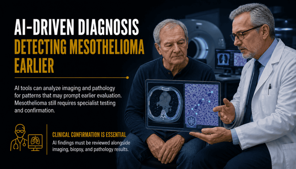
AI-Driven Diagnosis
Can artificial intelligence help detect mesothelioma earlier?
Artificial intelligence is being studied as a tool for analyzing medical images, digitized pathology slides and complex clinical data. In mesothelioma research, investigators hope AI can help identify suspicious patterns, measure irregular tumors and support specialist review.
AI does not independently diagnose mesothelioma or replace a representative tissue biopsy. Most mesothelioma-specific applications remain investigational, and a model that performs well in one dataset may not work equally well in another hospital, scanner, patient population or disease setting.
🤖 AI and Mesothelioma Diagnosis Overview
| AI Function | Possible Mesothelioma Research Use | What Human Experts Still Do |
|---|---|---|
| Detection | Flag pleural abnormalities or suspicious slide regions | Determine whether the finding is clinically meaningful |
| Segmentation | Outline tumor or fluid on imaging | Correct boundaries and interpret anatomy |
| Classification | Estimate which image or tissue pattern is present | Integrate morphology, stains, imaging and clinical history |
| Quantification | Measure tumor volume, texture or marker expression | Confirm measurement quality and relevance |
| Prediction | Study prognosis or possible treatment response | Explain uncertainty and make patient-specific decisions |
| Workflow support | Prioritize studies or retrieve similar cases | Maintain responsibility for diagnosis and follow-up |
The broader mesothelioma research overview explains how experimental tools move from laboratory development toward clinical validation. A research model should not be described as routine screening or diagnosis without evidence for that exact intended use.
🩻 AI in Medical Imaging
Mesothelioma can grow as irregular pleural thickening rather than a simple round mass. It may surround the lung, involve fissures or coexist with pleural fluid and scarring. These features create difficult detection and measurement problems for both automated systems and clinicians.
| Imaging Task | Potential AI Contribution | Important Limitation |
|---|---|---|
| Chest X-ray triage | Flag pleural fluid or another abnormal pattern for review | X-rays cannot reliably confirm or exclude mesothelioma |
| CT abnormality detection | Highlight pleural thickening, nodularity or volume change | Benign pleural disease and prior treatment can look similar |
| Tumor segmentation | Outline irregular pleural disease for measurement | Manual correction may be needed near lung, chest wall and fluid |
| PET/CT analysis | Quantify metabolic patterns or combine functional and anatomic data | Inflammation can produce uptake that resembles active tumor |
| MRI analysis | Study soft-tissue invasion or quantitative image features | Motion, acquisition differences and limited datasets affect performance |
| Longitudinal comparison | Align scans and estimate change over time | Different scanners, protocols and breathing position can distort comparisons |
AI may eventually make measurements faster or more reproducible, but it cannot determine malignancy from an image alone. Suspicious imaging normally leads to additional evaluation rather than a final mesothelioma diagnosis.
The site’s mesothelioma diagnosis overview explains how imaging fits with physical examination, procedures and pathology.
🔬 AI in Digital Pathology
Digital pathology converts glass slides into high-resolution images that can be reviewed on a computer and analyzed with software. AI research may help locate tumor-rich areas, count stained cells, quantify patterns or support classification.
| Pathology Application | Possible Benefit | Why Expert Review Remains Essential |
|---|---|---|
| Tumor-region detection | Direct attention to suspicious areas on a large slide | The model may overlook sparse or unusual tumor patterns |
| Cell-type classification | Study epithelioid, sarcomatoid and biphasic features | A small biopsy may not represent the entire tumor |
| Immunostain quantification | Measure staining intensity or the proportion of positive cells | Preanalytics, antibody selection and thresholds affect results |
| Mitotic or nuclear features | Quantify features associated with tumor behavior | Artifacts and tissue quality can create false measurements |
| Similarity search | Retrieve visually related reference cases | Similar appearance does not prove the same diagnosis |
| Quality control | Flag blurred areas, folds or incomplete scans | The original tissue and glass slide may still need reinspection |
Mesothelioma can resemble metastatic carcinoma, reactive mesothelial proliferation, sarcoma and other conditions. A pathologist must interpret morphology, invasion and an appropriate panel of immunostains. AI classification cannot compensate for inadequate tissue.
A specialist pathology second opinion remains valuable when the biopsy is small, findings conflict or a major treatment decision depends on the exact histology.
Questions After a Mesothelioma Diagnosis?
Our team can help you locate information and understand possible legal options related to asbestos exposure.
Call 800.291.0963 Submit Your Case
Live chat is available 24/7/365.
📊 Radiomics and Disease Measurement
Radiomics converts medical images into quantitative features such as shape, intensity and texture. Researchers may combine these features with clinical data to study diagnosis, disease extent, prognosis or treatment response.
| Research Goal | Example Output | Validation Question |
|---|---|---|
| Distinguish tissue patterns | Probability that a pleural finding belongs to a selected category | Was the model tested against appropriate benign and malignant mimics? |
| Estimate tumor burden | Automated volume or surface measurement | Does it remain accurate across scanners, sites and disease shapes? |
| Assess response | Change in volume, texture or metabolic features | Does the change predict a meaningful clinical outcome? |
| Predict prognosis | Risk category generated from images and clinical variables | Does it add value beyond stage, histology and performance status? |
| Plan local treatment | Automated contours for surgery or radiation planning | How often must specialists correct the proposed boundary? |
| Combine data types | Integrated imaging, pathology and molecular score | Are missing data, privacy and model transparency addressed? |
A model can be statistically accurate without improving patient care. Useful validation asks whether the tool changes a decision, reduces harmful delay, improves reproducibility or produces better outcomes without creating excessive false alarms.
Research measurements also should not be confused with clinical stage. The mesothelioma staging guide explains how disease extent is evaluated in practice.
🧬 AI, Biomarkers and Clinical Data
Machine-learning methods can search for patterns across laboratory values, tissue markers, genomic data, exposure history and medical records. These methods may help generate hypotheses or identify groups for further study.
| Data Source | Possible Research Use | Key Risk |
|---|---|---|
| Blood proteins | Study diagnostic, prognostic or monitoring signatures | Markers may be abnormal for reasons unrelated to mesothelioma |
| Genomic data | Explore inherited risk, tumor biology or trial eligibility | Association does not prove clinical usefulness |
| Electronic health records | Find patterns of symptoms, testing and diagnostic delay | Records may be incomplete, inconsistent or biased |
| Exposure histories | Study relationships between asbestos exposure and disease | Recall and documentation may be incomplete |
| Pathology and imaging together | Develop integrated diagnostic or prognostic models | Complex models can be difficult to interpret and reproduce |
| Longitudinal data | Model changes before and after treatment | Missing visits and treatment differences can distort results |
No blood biomarker or AI score currently replaces tissue confirmation in routine mesothelioma diagnosis. A model trained to distinguish known mesothelioma cases from healthy controls may perform much worse when faced with real-world patients who have other pleural diseases.
The site’s guide to mesothelioma biopsy explains why tissue architecture, invasion and immunohistochemistry remain central.
⚠️ Limitations, Bias and Clinical Validation
NCI notes that nonrepresentative training data can perpetuate medical bias and that further clinical trials are needed to validate AI applications. Rare cancers create added difficulty because datasets may be small, drawn from a few specialist centers or missing important comparison cases.
| Risk | How It Can Affect Results | Safeguard |
|---|---|---|
| Small dataset | The model memorizes local patterns rather than learning generalizable features | Independent external and prospective validation |
| Selection bias | Training cases do not represent the patients seen in practice | Diverse sites, demographics, disease stages and mimics |
| Data leakage | Information from the test set unintentionally influences training | Strict separation of patients, sites and time periods |
| Scanner or slide variation | Performance falls when equipment or preparation changes | Multivendor testing and monitoring after deployment |
| False positives | Benign findings trigger anxiety, scans or invasive procedures | Report specificity, predictive values and downstream consequences |
| False negatives | Disease is missed or falsely reassured | Human review, safety thresholds and clear fallback procedures |
| Automation bias | Clinicians over-trust a software suggestion | Training, explainability and documented human responsibility |
Accuracy alone is incomplete. Sensitivity, specificity, positive predictive value, negative predictive value, calibration and performance in clinically relevant subgroups all matter. Predictive values can change sharply when a rare disease is studied in a general population.
The FDA maintains a list of authorized AI-enabled medical devices, but inclusion applies only to the device’s cleared or approved intended use. It does not establish that every AI imaging product can detect mesothelioma.
🩺 What Mesothelioma Diagnosis Still Requires
Current diagnosis begins with clinical evaluation and appropriate imaging. When findings are suspicious, clinicians may drain fluid, obtain tissue and perform additional staging tests. Tissue is needed to classify the tumor and distinguish it from diseases with overlapping appearances.
| Diagnostic Element | Purpose | Role of AI Today |
|---|---|---|
| History and examination | Assess symptoms, health, prior disease and possible exposures | May organize information but cannot replace clinical judgment |
| Imaging | Locate abnormalities and guide procedures | May assist detection, segmentation or measurement in defined uses |
| Fluid analysis | Evaluate an effusion and sometimes identify malignant cells | Automated analysis is investigational and negative fluid does not exclude disease |
| Tissue biopsy | Show architecture, invasion and tumor morphology | AI may highlight or quantify areas on digitized slides |
| Immunohistochemistry | Support mesothelial lineage and exclude competing tumors | Software may quantify stains but cannot choose or interpret the panel alone |
| Molecular testing | Support malignancy or answer selected clinical questions | AI may integrate results but does not make every alteration actionable |
| Multidisciplinary review | Combine radiology, pathology and clinical findings | AI may support, not replace, accountable specialists |
Earlier diagnosis depends on more than software. Symptoms must be recognized, appropriate imaging ordered, abnormalities followed, adequate tissue obtained and pathology interpreted correctly. A faster algorithm cannot fix every delay elsewhere in the care pathway.
Seek prompt medical care for worsening shortness of breath, persistent chest or abdominal pain, unexplained fluid buildup, weight loss or another concerning symptom. Severe breathing difficulty, fainting or rapidly worsening chest pain requires urgent evaluation.
📋 Questions to Ask About an AI Diagnostic Tool
- What is the intended use? Ask whether the tool detects, measures, prioritizes or classifies—and for which patients.
- Is it mesothelioma-specific? A general lung or chest-imaging tool may not have been validated for pleural mesothelioma.
- Was it tested outside the development site? External validation helps reveal performance problems.
- What were the comparison cases? Relevant benign pleural disease and other cancers should be represented.
- How are false results handled? Ask what follow-up occurs after a positive, negative or uncertain output.
- Who makes the final decision? A qualified clinician should remain responsible for interpretation and care.
Authoritative Medical and Research Sources
- National Cancer Institute: Artificial Intelligence and Cancer
- U.S. Food and Drug Administration: AI-Enabled Medical Devices
- National Cancer Institute: Malignant Mesothelioma Treatment (PDQ), Health Professional Version
- Centers for Disease Control and Prevention: Mesothelioma Basics
- National Library of Medicine PubMed: Mesothelioma and Artificial Intelligence Research
- ClinicalTrials.gov: Mesothelioma Studies
❓ Frequently Asked Questions
Can AI diagnose mesothelioma?
AI can assist with selected imaging or pathology tasks, but it does not independently establish the diagnosis. Clinical evaluation, adequate tissue and expert pathology remain essential.
Can AI detect mesothelioma before symptoms begin?
This is a research goal, not an established routine capability. No AI result should be treated as proof that an asymptomatic person has or does not have mesothelioma.
Does AI replace a mesothelioma biopsy?
No. Imaging may show suspicious disease, but representative tissue is generally needed to assess morphology, invasion, immunostains and cell type.
What is radiomics?
Radiomics is the extraction of quantitative features such as shape, intensity and texture from medical images. Researchers study whether these features add clinically useful information.
Can AI tell whether a pleural abnormality is benign or malignant?
Research models may estimate probabilities, but benign disease, inflammation and other cancers can overlap with mesothelioma. Clinicians must confirm suspicious findings.
Are AI mesothelioma tools FDA approved?
The FDA lists many AI-enabled medical devices for defined intended uses. Patients should verify the exact device and indication; general AI authorization does not establish a mesothelioma-specific claim.
Can AI predict which mesothelioma treatment will work?
Researchers are studying treatment-response models, but no AI prediction guarantees benefit. Treatment decisions still depend on pathology, disease extent, health, evidence and patient goals.
What should I do if an AI report conflicts with my doctor?
Ask the clinician to explain the tool’s intended use, confidence and limitations. A specialist radiology or pathology review may help resolve a consequential disagreement.
Talk With Our Mesothelioma Support Team
If you or a loved one has been diagnosed with mesothelioma, we can help you find information and understand possible legal options related to asbestos exposure. There is no obligation.
Call 800.291.0963 Submit Your Case
Live chat is available 24/7/365.
Medical and legal disclaimer: This page provides general educational information and is not a substitute for advice from a licensed physician or attorney. AI outputs must be interpreted by qualified health professionals. Diagnosis, treatment, trial eligibility and legal rights depend on individual facts; contacting us does not create an attorney-client relationship.