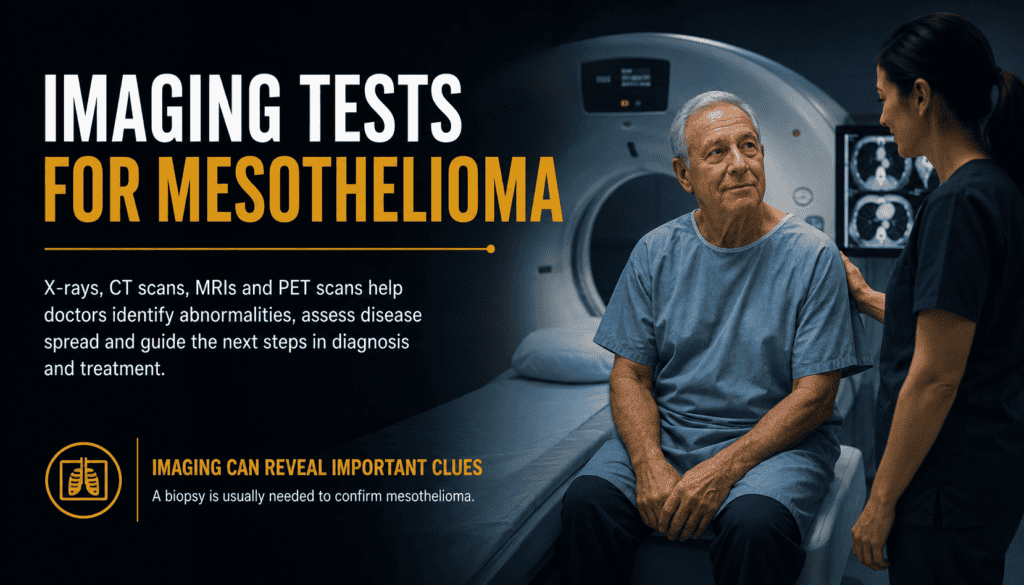
Imaging Tests for Mesothelioma
How X-rays, CT scans, PET scans, MRI and ultrasound help doctors investigate symptoms, map disease and plan the next step.
Imaging tests let clinicians see abnormalities in the chest or abdomen without surgery. They may reveal fluid, thickened tissue, a mass or possible spread. But a scan alone cannot establish a mesothelioma diagnosis: a clinician usually needs tissue examined by a pathologist. If you are just beginning an evaluation, see our mesothelioma diagnosis overview and symptom guide.
Urgent symptoms: New severe breathlessness, fainting, severe chest pain or rapidly worsening symptoms need prompt medical evaluation. Do not wait for a scheduled scan if you are struggling to breathe.
What imaging tests can—and cannot—tell you
Doctors choose scans based on symptoms, the suspected site of disease, previous findings and the question they need answered. A chest X-ray may start the investigation; CT provides detailed anatomy; PET assesses metabolic activity; MRI can clarify soft-tissue involvement. Ultrasound can help identify fluid and guide some procedures. Imaging is also used after diagnosis to assess disease extent and monitor treatment.
| Clinical question | How imaging may help | Important limitation |
|---|---|---|
| Why am I short of breath? | Look for pleural fluid, lung changes or a mass. | Several noncancer conditions can look similar. |
| Where is an abnormality? | Locate thickened pleura or peritoneum and adjacent structures. | Location does not establish cell type. |
| Has disease spread? | Evaluate lymph nodes and other areas as clinically indicated. | Small or microscopic deposits may not be visible. |
| Is treatment working? | Compare scans over time using consistent techniques. | Interpretation also depends on symptoms and the care plan. |
The mesothelioma stages guide explains how imaging findings contribute to staging alongside other evidence.
Chest X-rays: the first look
A chest X-ray is fast and commonly ordered for persistent cough, chest discomfort or breathlessness. It may show a pleural effusion (fluid around the lung), pleural thickening or other changes. A normal X-ray does not reliably exclude mesothelioma, and an abnormal one cannot identify the cancer by itself.
| Possible finding | What it means | Likely follow-up |
|---|---|---|
| Pleural effusion | Fluid is visible around a lung. | CT and possible fluid evaluation, depending on symptoms. |
| Pleural thickening | Lining appears thicker than expected. | CT for a more detailed assessment. |
| Pleural plaques | May reflect prior asbestos exposure. | Clinical review; plaques alone do not mean cancer. |
| No obvious abnormality | No clear finding on this image. | Further evaluation if symptoms or concern persist. |
Read about shortness of breath and fluid management if these are the symptoms that prompted imaging.
CT scans of the chest and abdomen
Computed tomography uses X-rays and a computer to create cross-sectional images. Chest CT can show the distribution of pleural thickening, masses, fluid and nearby structures more clearly than a plain X-ray. Abdominal CT may be appropriate when symptoms or findings suggest peritoneal disease. Contrast material may be used to improve visibility, but the radiology team decides whether it is appropriate.
| CT feature | Why it matters | What to ask |
|---|---|---|
| Pleural rind or nodules | Helps map suspicious tissue around the lung. | Where is the thickening and how extensive is it? |
| Pleural fluid | Shows the amount and location of fluid. | Could drainage help my symptoms or testing? |
| Chest wall or diaphragm contact | May prompt closer evaluation of local involvement. | Would MRI clarify this finding? |
| Abdominal nodules or fluid | May suggest a peritoneal process. | Do I need abdominal imaging or a biopsy? |
| Before CT | Practical point |
|---|---|
| Contrast history | Tell the team about prior contrast reactions and kidney problems. |
| Medications | Follow the imaging center’s specific medication instructions. |
| Pregnancy possibility | Tell the team before radiation-based imaging. |
| Prior images | Bring outside scans or arrange image transfer for comparison. |
CT findings help guide a mesothelioma biopsy but cannot replace the tissue diagnosis.
PET and PET/CT scans
Positron emission tomography often uses a small amount of radioactive glucose tracer called FDG. Areas that use more tracer may appear more active. Combined PET/CT overlays metabolic information on detailed anatomy. Clinicians may use it after a suspected or established diagnosis to evaluate possible spread or investigate a suspicious area.
Important: Increased tracer uptake is not proof of cancer. Infection and inflammation can also be active, and some cancers or small deposits may be missed. Results must be interpreted with CT, clinical history and pathology.
| PET/CT question | Potential contribution | Caution |
|---|---|---|
| Is this thickening metabolically active? | Identifies regions of higher FDG uptake. | Inflammation can also cause uptake. |
| Are lymph nodes suspicious? | Highlights nodes that merit closer review. | Biopsy may be needed to establish involvement. |
| Is there disease elsewhere? | Surveys additional areas when indicated. | Very small disease can be below scan resolution. |
| Should a site be sampled? | May help identify a target. | Sampling decision depends on accessibility and safety. |
| Preparation issue | Why it matters |
|---|---|
| Fasting instructions | Food and glucose levels can affect FDG distribution. |
| Diabetes | Ask for individualized medication and appointment instructions. |
| Recent infection or surgery | Tell the team; inflammation can affect interpretation. |
| Pregnancy or breastfeeding | Notify the imaging service for tailored guidance. |
MRI and ultrasound
MRI uses magnets and radio waves rather than ionizing radiation. In selected cases it can better define soft tissues, including possible involvement of the diaphragm, chest wall or nearby structures. MRI is not necessary for every patient. Tell the team about implanted devices, metal fragments, kidney disease or difficulty tolerating enclosed spaces before the appointment.
Ultrasound uses sound waves. It can help locate pleural or abdominal fluid and guide certain fluid-drainage procedures or biopsies. An echocardiogram, a specialized ultrasound of the heart, may be used when fluid around the heart is suspected.
| Test | Best-known role | Main limitation |
|---|---|---|
| MRI | Detailed soft-tissue assessment in selected situations. | Longer exam; device and contrast considerations. |
| Chest ultrasound | Identify fluid and assist safe procedure planning. | Not a comprehensive cancer-staging test. |
| Abdominal ultrasound | Assess ascites and guide drainage or sampling. | May not characterize all peritoneal deposits. |
| Echocardiogram | Assess heart function and possible pericardial fluid. | Does not diagnose pleural mesothelioma. |
Different imaging pathways may be used for pleural mesothelioma and peritoneal mesothelioma.
Understanding your imaging report
A radiology report usually describes the technique, findings, comparison with earlier studies and the radiologist’s impression. Terms such as “suspicious,” “indeterminate” or “cannot exclude” express uncertainty—not a confirmed diagnosis. Ask the ordering clinician to explain how the report fits your symptoms and whether additional testing is warranted.
| Report term | Plain-language meaning | Follow-up question |
|---|---|---|
| Pleural effusion | Fluid between lung and chest wall. | Is it affecting breathing, and should it be sampled? |
| Pleural thickening | Lining appears thicker than expected. | Is it focal or diffuse, and has it changed? |
| Nodule or mass | A localized abnormal area. | What is the recommended next test? |
| Lymphadenopathy | Enlarged lymph nodes. | Is tissue sampling needed? |
| Indeterminate | The scan cannot identify the cause confidently. | What evidence would resolve the uncertainty? |
| Comparison | Why clinicians care |
|---|---|
| Earlier baseline scan | Shows whether a finding is longstanding. |
| Same imaging method | Can make changes easier to interpret. |
| Measurements and symptoms | Both matter when assessing response. |
| Radiologist impression | Summarizes findings but does not replace pathology. |
What happens after an abnormal scan
If imaging raises concern for mesothelioma, a specialist may recommend tissue sampling. CT or ultrasound can guide a needle toward an accessible abnormality; thoracoscopy or laparoscopy may be used when a larger or more representative specimen is needed. A pathologist examines the sample and may use immunohistochemical markers to distinguish mesothelioma from other cancers.
Fluid cytology can provide useful information, but a negative fluid result does not necessarily rule out mesothelioma. A specialist may recommend further tissue sampling when suspicion remains. Blood biomarkers are not a substitute for biopsy.
| Next step | Purpose |
|---|---|
| Review scan with specialist | Identify the most informative and safest sampling approach. |
| Obtain tissue | Allow microscopic examination and additional testing. |
| Pathology review | Determine whether mesothelioma is present and identify subtype. |
| Stage and plan care | Combine pathology, imaging and clinical findings. |
See how mesothelioma biopsies work, blood tests and biomarkers and mesothelioma treatments for the next parts of the care pathway.
Preparing for scans and follow-up
Ask what question each scan is intended to answer. Confirm whether contrast or tracer will be used, whether you need to fast, and how you will receive results. Keep copies of the radiology report and the actual image files; specialists may need both. If symptoms worsen while you are waiting, contact your care team rather than assuming an appointment can wait.
| Question for your team | Why it is useful |
|---|---|
| What are we looking for? | Clarifies the purpose of the test. |
| Do I need contrast? | Allows a discussion of benefits and precautions. |
| Will I need a biopsy? | Sets expectations about diagnostic certainty. |
| When will results be discussed? | Creates a clear follow-up plan. |
| Should previous images be compared? | Helps evaluate change over time. |
| After the scan | Action |
|---|---|
| Get the report | Request a copy for your records. |
| Ask about unclear language | Discuss findings with the ordering clinician. |
| Confirm the next appointment | Know who coordinates further testing. |
| Track symptoms | Report new or worsening problems promptly. |
Frequently asked questions
Can an X-ray diagnose mesothelioma?
No. It may show fluid or thickening that prompts further tests, but tissue examination is generally required for diagnosis.
Is CT better than a chest X-ray?
CT provides more detailed cross-sectional anatomy. The tests answer different questions; a clinician chooses based on the situation.
Does a PET scan prove a mass is cancer?
No. Infection and inflammation may also cause increased tracer uptake, and some cancer may not appear prominently.
Why might my doctor order MRI after CT?
MRI may clarify possible involvement of the diaphragm, chest wall or other soft tissues when that information would affect planning.
Can scans tell whether mesothelioma has spread?
They help assess extent, but imaging may miss microscopic disease. Other tests or biopsies may be needed.
Can a normal scan rule out mesothelioma?
Not reliably in every circumstance. Persistent concerning symptoms and asbestos history deserve clinical assessment.
What if fluid testing is negative but CT remains suspicious?
A specialist may recommend a tissue biopsy or additional evaluation because fluid testing can miss mesothelioma.
Should I request every scan?
Not necessarily. More imaging is not always more useful. Ask which test will change diagnosis, staging or treatment decisions.
Confirmed Medical Sources
These sources support the diagnostic roles and limitations described above:
Get Help After a Mesothelioma Diagnosis
Call 800.291.0963. Live chat is available 24/7, 365 days a year. Submit Your Case.
Contacting us does not guarantee eligibility, compensation or a particular outcome. Medical concerns should be addressed with your treating team.
⚕️ Legal & Medical Information Disclaimer
This article provides general information, not personalized medical or legal advice. Imaging and biopsy decisions belong to qualified clinicians; consult a licensed attorney about legal questions.