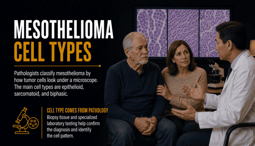
Mesothelioma Cell Types
Understanding epithelioid, sarcomatoid and biphasic histology
Mesothelioma cell type describes how tumor cells look and grow under a microscope. The three principal histologic types are epithelioid, sarcomatoid and biphasic. A pathology report may also identify a more specific pattern, grade or molecular finding that helps confirm the diagnosis.
Cell type matters because it contributes to prognosis and may influence surgery, systemic therapy and clinical-trial decisions. It is only one part of the picture: tumor location and stage, lymph-node involvement, overall health, treatment goals and specialist review also shape the care plan.
🧬 Mesothelioma Cell Types Overview
Cell type is also called histology or histologic subtype. A pathologist determines it by examining tissue from a tumor and using additional tests to confirm that the cells are malignant mesothelial cells rather than another cancer or a benign process.
Cell type and tumor location answer different questions. Location identifies where the cancer began—most often the pleura around the lungs or the peritoneum in the abdomen. Histology identifies the tumor’s microscopic appearance. A person may therefore have pleural epithelioid mesothelioma, peritoneal biphasic mesothelioma or another location-and-cell-type combination.
The National Cancer Institute describes malignant mesothelioma as containing epithelial elements, spindle-shaped sarcomatoid cells or both. These categories are:
- Epithelioid: tumor cells grow in epithelial-like arrangements and may form tubules, papillae, sheets or other patterns.
- Sarcomatoid: tumor cells are often elongated or spindle-shaped and grow in less cohesive arrangements.
- Biphasic: the tumor contains both epithelioid and sarcomatoid components.
⚖️ Epithelioid, Sarcomatoid and Biphasic Compared
| Feature | Epithelioid | Sarcomatoid | Biphasic |
|---|---|---|---|
| Basic appearance | Epithelial-like, often cohesive cells | Spindle-shaped or elongated cells | Both epithelioid and sarcomatoid areas |
| Relative frequency | Most common histologic type | Least common of the three principal types | Mixed histology found between the other two in frequency |
| Typical behavior | Often more cohesive and generally associated with a more favorable outlook | Often more infiltrative and generally associated with a less favorable outlook | Behavior varies with tumor biology and the balance and distribution of both components |
| Diagnostic challenge | May resemble adenocarcinoma or another epithelioid tumor | May resemble sarcoma, reactive fibrosis or sarcomatoid carcinoma | A small sample may capture only one component |
| Treatment relevance | May be one factor supporting surgical evaluation in otherwise suitable patients | Often limits the role of major surgery and can influence systemic-treatment discussions | Requires individualized assessment; the sarcomatoid component is clinically important |
These patterns describe broad clinical tendencies, not a guaranteed outcome for an individual. Two tumors with the same label can differ in stage, molecular changes, treatment response and growth rate.
🔵 Epithelioid Mesothelioma
Epithelioid mesothelioma is the most common principal histologic type. NCI’s professional treatment summary states that epithelioid tumors account for approximately 60% of mesothelioma diagnoses, although proportions can differ among studies, locations and classification methods.
Under the microscope, epithelioid cells tend to be more cohesive than sarcomatoid cells. They can form several recognized architectural patterns. Some patterns and cytologic features may carry prognostic information, but the complete report and clinical setting matter more than a pattern name viewed alone.
| Epithelioid Feature | What It May Mean | Important Limitation |
|---|---|---|
| Tubulopapillary, trabecular or solid architecture | Describes how tumor cells are arranged | Architecture alone does not establish malignancy or determine treatment |
| Nuclear grade and mitotic activity | May help describe how abnormal and actively dividing the cells appear | Grading practices depend on tumor site and pathology standards |
| Necrosis | Dead tumor tissue may provide additional prognostic information | Its meaning must be interpreted with grade, stage and sampling quality |
| Mesothelial immunoprofile | Supports mesothelial differentiation | No single immunostain proves every case |
Epithelioid histology is generally associated with better outcomes than sarcomatoid or biphasic histology. It may also be considered when specialists evaluate whether a patient could benefit from a multimodality plan that includes surgery. However, epithelioid disease can still be advanced, unresectable or resistant to treatment.
🔶 Sarcomatoid Mesothelioma
Sarcomatoid mesothelioma is composed predominantly of spindle-shaped malignant cells. These cells may be widely separated, arranged in fascicles or embedded in dense connective tissue. The tumor can be difficult to distinguish from true sarcoma, sarcomatoid carcinoma and noncancerous fibrous pleural disease.
Desmoplastic mesothelioma is a recognized sarcomatoid subtype characterized by dense collagen with relatively inconspicuous malignant cells. Because it may resemble benign pleural fibrosis, proving invasion and using an appropriate immunohistochemical and molecular workup can be especially important.
| Challenge | Why It Matters | Possible Pathology Approach |
|---|---|---|
| Spindle-cell appearance | Several cancers and sarcomas can look similar | Use a differential-diagnosis panel rather than one marker |
| Dense fibrosis | Malignant cells may be sparse or subtle | Look for tissue invasion and loss of certain tumor-suppressor markers when appropriate |
| Small biopsy | The sample may not represent the whole tumor | Correlate with imaging and consider additional tissue if results remain uncertain |
| Limited treatment sensitivity | Some conventional approaches may be less effective | Discuss systemic therapy and clinical trials with a specialist |
Sarcomatoid histology is generally associated with a less favorable prognosis than epithelioid histology. It often reduces enthusiasm for aggressive surgery, but it does not mean that supportive care is the only option. Treatment may include immunotherapy, chemotherapy, radiation for specific symptoms or sites, and a clinical trial, depending on location and the patient’s condition.
🟡 Biphasic Mesothelioma
Biphasic mesothelioma, sometimes called mixed mesothelioma, contains both epithelioid and sarcomatoid components. A definitive classification depends on adequate tissue sampling because the two components may not be evenly distributed throughout a tumor.
A core biopsy taken from an epithelioid area may miss a nearby sarcomatoid component, while a sample from a spindle-cell area may not show the epithelial portion. This is one reason a pathology classification can change after a larger biopsy, surgery or expert review. A changed diagnosis does not necessarily mean that the first pathologist made an obvious error; the additional specimen may simply reveal tumor heterogeneity.
Why the Mixture Matters
- The sarcomatoid component can influence tumor behavior and the expected benefit-risk balance of surgery.
- A larger specimen may provide a more representative estimate of both components.
- The percentage of each component should not be used as a stand-alone survival calculator.
- Treatment still depends on site, stage, resectability, performance status and patient preferences.
Biphasic outcomes are often described as intermediate between epithelioid and sarcomatoid disease, but that shorthand can hide wide individual variation. The pathology report should be reviewed alongside the complete clinical record.
Get Help After a Mesothelioma Diagnosis
Our team can help you locate information and understand possible legal options related to asbestos exposure.
Call 800.291.0963 Submit Your Case
Live chat is available 24/7/365.
🔬 How Biopsy and Pathology Identify Cell Type
Imaging can show pleural thickening, abdominal nodules, a mass or fluid, but it cannot reliably identify mesothelioma cell type. A tissue mesothelioma biopsy allows a pathologist to examine tumor architecture, cellular details and invasion.
| Diagnostic Step | Purpose | What Patients Should Know |
|---|---|---|
| Review of tissue architecture | Evaluates epithelioid, spindle-cell or mixed growth | Larger samples may show features absent from a small biopsy |
| Immunohistochemistry | Uses antibodies to assess mesothelial and alternative tumor markers | Panels are chosen for the specific differential diagnosis |
| Evidence of malignancy | Distinguishes invasive cancer from reactive mesothelial proliferation | Invasion, BAP1 loss, MTAP loss or CDKN2A deletion may help in appropriate settings |
| Clinical-radiologic correlation | Compares pathology with tumor location and imaging | A result that conflicts with the clinical pattern may require review |
| Expert second review | Reassesses rare or difficult cases | Original slides, tissue blocks and reports may be requested |
Common Immunohistochemical Markers
Depending on the suspected diagnosis, a pathologist may use positive mesothelial markers such as calretinin, WT1, D2-40 and cytokeratin 5/6, along with markers intended to exclude adenocarcinoma or another malignancy. Marker performance differs by cell type and tumor site. A panel is more reliable than relying on a single stain.
Loss of nuclear BAP1 staining or loss of MTAP expression can support malignancy in a mesothelial proliferation, and testing for CDKN2A deletion may be useful in selected cases. A retained marker does not automatically rule out mesothelioma. These tests must be interpreted by a qualified pathologist in context.
💊 How Cell Type Affects Treatment and Prognosis
Histology is an important prognostic factor, but it does not independently dictate treatment. Specialists combine it with tumor site, stage or distribution, resectability, lymph-node status, symptoms, organ function, performance status and the patient’s goals.
| Decision Area | How Cell Type May Contribute | Other Essential Factors |
|---|---|---|
| Surgery | Epithelioid histology is more often associated with consideration of tumor-directed surgery; sarcomatoid histology may weigh against it | Stage, resectability, nodal disease, heart-lung function, age and operative risk |
| Systemic therapy | Histology may inform expected response and regimen discussion | Pleural versus peritoneal location, prior treatment, approvals, contraindications and patient preference |
| Radiation | Histology is one consideration but usually does not determine use by itself | Treatment field, nearby organs, symptoms and prior radiation |
| Clinical trials | Some studies enroll or stratify patients by histology or molecular findings | Eligibility rules, location, stage, laboratory results and previous therapy |
| Prognosis | Epithelioid disease generally has a more favorable population-level outlook than nonepithelioid disease | Individual outcomes vary and cannot be predicted from histology alone |
For unresectable malignant pleural mesothelioma, treatment choices can include immunotherapy, chemotherapy or other approved combinations. Evidence from modern trials shows why histology can matter in subgroup discussions, but a subgroup result is not a promise of benefit for one patient. Peritoneal mesothelioma has a different evidence base and may involve cytoreductive surgery with HIPEC for selected patients.
Explore the site’s mesothelioma treatment overview and mesothelioma prognosis information. Avoid interpreting a single survival statistic without knowing whether the study population matches the tumor location, stage, cell type, treatment era and patient characteristics.
📋 Questions for the Pathologist or Oncology Team
Patients do not need to interpret a pathology report alone. Useful questions include:
- Was the diagnosis based on fluid, a needle biopsy, thoracoscopy, laparoscopy or a surgical specimen?
- What is the exact histologic type, and was a more specific pattern or grade reported?
- Was the sample large enough to assess a possible biphasic tumor?
- Which immunohistochemical stains were positive and negative?
- Was BAP1, MTAP or CDKN2A testing performed, and what does the result mean in this case?
- Which alternative diagnoses were considered and excluded?
- Would review by a pathologist experienced in mesothelioma change treatment planning?
- How do location, stage and cell type combine to affect the recommended treatment?
Patients seeking a review should ask how to transfer pathology slides, paraffin blocks, imaging and procedure notes. Preserve copies of reports and confirm that the reviewing center has received adequate material before an appointment.
Authoritative Medical Sources
- National Cancer Institute: Malignant Mesothelioma Treatment (PDQ), Health Professional Version
- National Cancer Institute: Mesothelioma
- College of American Pathologists: Pleural Mesothelioma Resection Protocol
- U.S. Food and Drug Administration: Nivolumab Plus Ipilimumab Approval for Mesothelioma
- Centers for Disease Control and Prevention: Mesothelioma Basics
- ClinicalTrials.gov: Mesothelioma Studies
❓ Frequently Asked Questions
What are the three main mesothelioma cell types?
The three principal histologic types are epithelioid, sarcomatoid and biphasic. Biphasic tumors contain both epithelioid and sarcomatoid components.
Which mesothelioma cell type is most common?
Epithelioid is the most common principal type. The National Cancer Institute’s professional summary estimates that it accounts for approximately 60% of mesothelioma diagnoses, although reported proportions vary.
Which cell type has the best prognosis?
Epithelioid histology is generally associated with a more favorable population-level prognosis than sarcomatoid or biphasic histology. An individual’s outlook still depends on location, stage, treatment options, health and response.
Can a mesothelioma cell-type diagnosis change?
Yes. A larger or better-targeted specimen can reveal features that were not present in the initial sample. Expert review may also refine the diagnosis after additional immunohistochemical or molecular testing.
Can a blood test identify mesothelioma cell type?
No routine blood test can replace tissue pathology for classifying cell type. Blood biomarkers may contribute to research or clinical assessment, but a biopsy remains central to diagnosis.
Can imaging distinguish epithelioid from sarcomatoid mesothelioma?
Imaging shows tumor location and extent but cannot reliably determine histology. Cell type is established by examining tissue under a microscope with supporting laboratory tests.
Does sarcomatoid mesothelioma mean there are no treatment options?
No. Sarcomatoid disease is challenging and may not be suitable for aggressive surgery, but systemic therapy, radiation for selected problems, symptom management and clinical trials may still be considered.
Should every mesothelioma pathology report receive a second opinion?
Not every case requires one, but review by an experienced mesothelioma pathologist is reasonable when tissue is limited, findings conflict, the diagnosis is unusual or a major treatment decision depends on histology.
Talk With Our Mesothelioma Support Team
If you or a loved one has been diagnosed with any mesothelioma cell type, we can help you find information and understand possible legal options related to asbestos exposure. There is no obligation.
Call 800.291.0963 Submit Your Case
Live chat is available 24/7/365.
Medical and legal disclaimer: This page provides general educational information and is not a substitute for advice from a licensed physician or attorney. Diagnosis and treatment decisions must be made with qualified health professionals. Case eligibility and legal rights depend on individual facts and applicable law; contacting us does not create an attorney-client relationship.