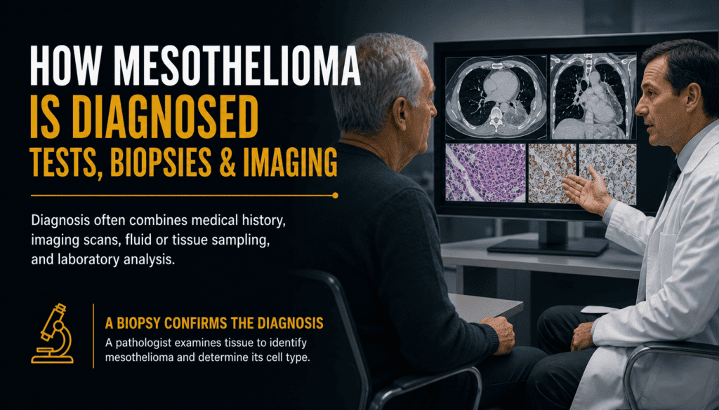
How Mesothelioma Is Diagnosed: Tests, Biopsies and Imaging
A patient-friendly guide to the diagnostic process, what each test can reveal, and why a tissue diagnosis often matters.
Mesothelioma diagnosis usually involves several steps—not one definitive scan or blood test. A clinician reviews symptoms and exposure history, orders imaging to locate abnormalities, and often obtains tissue for a pathologist to examine. The results help distinguish mesothelioma from other conditions and guide decisions about treatment. This guide explains the process without assuming that any particular symptom or test result proves cancer.
Seek urgent medical attention for severe or rapidly worsening shortness of breath, new severe chest pain, fainting, or other sudden serious symptoms. Contact your healthcare team promptly about persistent or worsening symptoms.
Diagnosis at a Glance
A suspicious imaging result starts a clinical investigation; it does not establish a diagnosis by itself. The sequence varies by the location of the abnormality, the patient’s health and whether previous samples were adequate. The mesothelioma diagnosis overview provides a broader introduction.
| Step | Main purpose | What it cannot establish alone |
|---|---|---|
| History and exam | Identify symptoms, risk factors and physical findings | Whether cancer is present |
| Imaging | Locate abnormal tissue, fluid and possible spread | Exact cancer type |
| Fluid sampling | Evaluate an effusion and sometimes relieve symptoms | A reliable exclusion when negative |
| Tissue biopsy | Obtain tissue for laboratory evaluation | Complete stage in every case |
| Pathology review | Identify cancer and subtype using microscopic and ancillary testing | The entire treatment plan without clinical context |
Doctors may change the order of tests if a patient needs urgent relief of fluid buildup, cannot safely undergo a procedure or already has a diagnostic specimen. Ask what question each proposed test is intended to answer.
Symptoms and Exposure History
A clinician may ask about chest discomfort, breathlessness, persistent cough, abdominal swelling, appetite changes, weight loss and how symptoms have evolved. These symptoms are not specific to mesothelioma. A physical exam can identify clues such as reduced breath sounds or abdominal distention. See mesothelioma symptoms for symptom-specific explanations.
Provide a chronological work, military, household and building-renovation history, including possible asbestos contact and dates when it occurred. An absence of remembered exposure does not automatically exclude mesothelioma; likewise, a history of exposure does not establish that a current symptom is cancer.
| Information to bring | Why it matters |
|---|---|
| Symptom timeline | Shows onset, persistence and progression |
| Prior chest or abdominal scans | Allows comparison with older findings |
| Occupational and military history | Identifies potential asbestos exposure context |
| Previous fluid or biopsy reports | Prevents duplication and highlights inconclusive results |
| Current medicines and allergies | Helps plan contrast imaging and procedures |
Tell the team about blood thinners, kidney disease, implanted devices, prior reactions to contrast and any condition that may affect sedation or biopsy safety.
Imaging Tests
Imaging can reveal pleural thickening, fluid around the lung, abdominal changes or other abnormalities and can guide a biopsy. The best test depends on the suspected disease location and the clinical question. Read the focused guide to imaging tests for mesothelioma.
| Test | Common diagnostic role | Important limitation |
|---|---|---|
| Chest X-ray | Initial look at fluid or chest abnormalities | Can miss subtle disease and cannot confirm cancer |
| CT scan | Detailed view of chest or abdomen; helps target biopsy | Appearance can overlap with other diseases |
| PET/CT | May help assess suspicious sites and extent in selected patients | Inflammation can also show uptake |
| MRI | Selected questions about soft-tissue involvement | Not required for every patient |
| Ultrasound | Locate fluid and help guide a sampling procedure | Cannot identify tumor subtype |
A radiology report may use terms such as “pleural effusion,” “pleural thickening,” “nodularity” or “suspicious lesion.” These describe observations or concern—not a final microscopic diagnosis. A normal or nonspecific X-ray does not necessarily end the workup when symptoms and clinical suspicion persist. See what mesothelioma X-rays can show.
Fluid Testing and Its Limits
Fluid can accumulate around the lungs (pleural effusion) or in the abdomen (ascites). Clinicians may remove some with a needle to evaluate its cause and, when appropriate, ease discomfort. The fluid is sent for cytology, in which a pathologist examines cells. The procedure is called thoracentesis for pleural fluid and paracentesis for abdominal fluid.
| Finding | Meaning | Possible next question |
|---|---|---|
| Cancer cells identified | Raises or establishes evidence of malignancy depending on analysis | Is there enough material to classify the cancer? |
| No malignant cells identified | Cancer cells were not seen in this sample | Is tissue biopsy still needed? |
| Insufficient sample | The lab could not answer the question | Can a different sampling approach help? |
A negative fluid cytology result does not rule out mesothelioma. It may be difficult to distinguish mesothelioma from other cancers in fluid alone, and a tissue sample may be needed for a reliable diagnosis and subtype. Fluid drainage can be a separate symptom-relief decision even when the diagnosis remains uncertain. Explore mesothelioma fluid management.
Biopsy Procedures
A biopsy removes cells or tissue for microscopic examination. For many patients, a tissue biopsy is needed to establish the diagnosis and distinguish mesothelioma from a different malignancy. The clinician chooses a technique based on tumor location, sample needs and procedure risks. Review thoracoscopy and laparoscopy for more detail.
| Procedure | How tissue is obtained | When it may be considered |
|---|---|---|
| Image-guided core needle biopsy | Needle targets an abnormal area with imaging assistance | Accessible lesion with an acceptable safety profile |
| Thoracoscopy | Camera enters chest through small incision(s) | Direct visualization and multiple pleural samples |
| Laparoscopy | Camera enters abdomen through small incision(s) | Suspicious peritoneal disease |
| Open surgical biopsy | Surgeon obtains tissue through an incision | When other approaches do not yield adequate tissue |
Ask whether the planned approach is likely to provide enough tissue for both diagnosis and cell-type classification. Possible complications vary by procedure and may include bleeding, infection, anesthesia-related problems or pneumothorax with chest biopsy. Your team should explain your individual risks, whether to hold medicines and how results will be communicated.
Pathology and Cell Types
A pathologist examines the sample and may order immunohistochemistry, which uses marker patterns to help identify mesothelioma and distinguish it from cancers that can look similar. No single stain is interpreted in isolation: the marker panel, tissue appearance and clinical findings are considered together. A difficult case may warrant review by a pathologist experienced with mesothelioma.
| Pathology term | What it describes | Why it matters |
|---|---|---|
| Epithelioid | A mesothelioma cell type | Can influence treatment discussion and prognosis |
| Sarcomatoid | A different cell pattern | Can influence treatment discussion and prognosis |
| Biphasic | Both epithelioid and sarcomatoid components | Adequate sampling helps characterize the mixture |
| Immunohistochemistry | Laboratory marker testing on tissue | Helps distinguish mesothelioma from mimics |
| Inconclusive / insufficient | Sample does not answer the diagnostic question | May prompt expert review or repeat biopsy |
Request a copy of the final pathology report, including any addendum. Ask whether the diagnosis and subtype are definitive, which findings support them, and whether an outside pathology review would be useful. For further context, see getting a mesothelioma second opinion.
Staging and Additional Tests
Diagnosis answers “What is this disease?” Staging asks “How extensive is it?” Additional imaging, lymph-node sampling or other procedures may be used after diagnosis to help plan care. Formal TNM stage groupings are established for pleural mesothelioma; clinicians evaluate peritoneal disease extent differently rather than automatically applying the same stage I–IV chart.
| Assessment | What it helps determine |
|---|---|
| CT and selected PET/CT or MRI | Anatomic extent and suspected involvement of other sites |
| Lymph-node sampling when indicated | Whether sampled nodes contain cancer |
| Pulmonary function tests | Lung function and suitability for certain procedures |
| General health assessment | Treatment tolerance and supportive-care needs |
Read mesothelioma stages and pulmonary function tests. A stage is one part of the clinical picture; the patient’s overall health, histology, treatment goals and preferences also matter.
What to Do After Results
Ask the diagnosing team for a clear explanation of what is confirmed, what remains uncertain and what happens next. A specialist may review the pathology slides, original scan images and procedural reports—not just a summary letter. If results conflict or a biopsy is nondiagnostic, ask what additional evaluation is appropriate rather than assuming the absence of cancer.
| Question to ask | Why it helps |
|---|---|
| Is mesothelioma confirmed or only suspected? | Separates a working diagnosis from a final result |
| What exact site and cell type were identified? | Clarifies the pathology report |
| Were the samples sufficient? | Identifies whether further tissue is needed |
| Has disease extent been evaluated? | Connects diagnosis with treatment planning |
| Should a mesothelioma specialist review the case? | Supports a considered second opinion |
| When will I receive reports and follow-up? | Prevents gaps in communication |
Keep a folder of imaging reports and image files, pathology reports and slides availability, procedure notes, medication lists and contact details. See articles for people diagnosed with mesothelioma and mesothelioma treatment information.
Confirmed medical sources
Frequently Asked Questions
Can an X-ray diagnose mesothelioma?
No. An X-ray may show fluid or other abnormalities, but imaging cannot establish the exact cancer type. A clinician may recommend CT imaging and tissue sampling.
Is a CT scan enough to confirm mesothelioma?
No. CT can identify suspicious areas and help guide a biopsy, but a pathological examination is generally needed for confirmation.
What is the most important test for a definite diagnosis?
A biopsy providing adequate tissue for examination and appropriate laboratory testing is often essential. The best sampling method depends on the location and individual circumstances.
Does a negative pleural fluid test rule out mesothelioma?
No. Cytology can miss mesothelioma or provide insufficient information. Doctors may recommend tissue biopsy when suspicion remains.
How are mesothelioma and lung cancer distinguished?
Pathologists evaluate tissue appearance and use panels of immunohistochemical markers together with the clinical and imaging findings.
Why might I need another biopsy?
An initial sample may be too small, may miss the abnormal area or may not permit reliable cell-type classification. Your team should explain why more tissue is necessary.
Do blood biomarkers confirm mesothelioma?
No. Biomarker results do not replace tissue-based diagnosis. Ask your clinician what any ordered blood test is intended to assess.
Should I get a second opinion after diagnosis?
A second opinion can help confirm a complex pathology diagnosis and clarify treatment options. Request copies of pathology reports, slides and imaging for the reviewing team.
Get Help After a Mesothelioma Diagnosis
Call 800.291.0963 to discuss your questions. Live chat is available 24/7, 365 days a year. Submit Your Case using the form below.
Submitting information does not guarantee compensation, legal representation or any particular outcome. Medical diagnosis and treatment decisions belong with qualified healthcare professionals.
⚕️ Legal & Medical Information Disclaimer
This article provides general educational information, not individualized medical or legal advice. Seek guidance from a qualified clinician for diagnosis or treatment and a licensed attorney for legal questions.