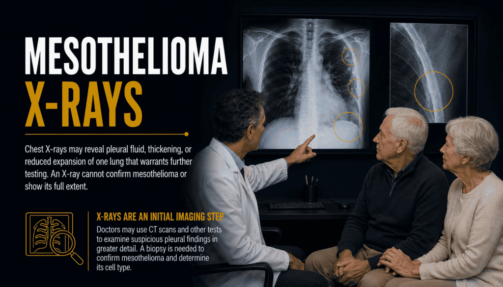
Mesothelioma X-Rays
What chest X-rays may reveal, why they cannot diagnose mesothelioma, and how doctors decide what to do next.
A chest X-ray is often an early step when someone has chest discomfort, a persistent cough, or trouble breathing. It can show changes that warrant more investigation, but it cannot establish whether those changes are mesothelioma. Doctors consider symptoms, asbestos exposure history, more detailed imaging, and often a tissue biopsy together.
Key takeaway: An abnormal X-ray is not a cancer diagnosis, and a normal X-ray does not rule out mesothelioma. If symptoms persist, ask your clinician what follow-up is appropriate.
What Is a Mesothelioma Chest X-Ray?
A chest radiograph uses a small amount of ionizing radiation to create a two-dimensional image of the chest. It is quick and widely available. A clinician may request it to investigate breathlessness, cough, chest pain, or an examination finding. The image can suggest an issue in the lung or its surrounding lining, but the picture does not identify the exact cell type.
Pleural mesothelioma develops in the lining around the lungs. Because this lining is thin and overlaps other chest structures on a flat image, early disease can be difficult to recognize on an X-ray. Learn about the broader mesothelioma diagnosis process and the role of different imaging tests.
| Term | Plain-language meaning |
|---|---|
| Chest X-ray | A flat image of the chest made with X-rays. |
| Pleura | The thin tissue covering the lungs and lining the inside of the chest. |
| Pleural effusion | Fluid collecting between the layers of pleura. |
| Pleural thickening | A lining that appears thicker than expected. |
| CT scan | A more detailed series of cross-sectional X-ray images. |
The National Cancer Institute lists chest X-ray among tests used when evaluating suspected mesothelioma; it also describes CT and biopsy as part of the diagnostic process. An X-ray is therefore a starting point, not a stand-alone answer.
What Can an X-Ray Show?
Radiologists look for patterns that may explain symptoms or justify further testing. Some findings can occur with mesothelioma, but they are not specific to it: infections, heart conditions, prior asbestos-related changes, and other cancers can produce overlapping appearances.
| Possible finding | What it may suggest | Why follow-up matters |
|---|---|---|
| Pleural effusion | Fluid around a lung | The cause may be cancerous or noncancerous. |
| Pleural thickening | An abnormal-looking lung lining | CT can define its location and shape more clearly. |
| Pleural calcifications or plaques | Possible prior asbestos-related pleural change | Plaques are not themselves a diagnosis of mesothelioma. |
| Reduced lung expansion or opacity | Fluid, lung disease, or another process | The image alone cannot reliably identify the cause. |
| A visible mass | An abnormal area requiring evaluation | Additional imaging and sometimes tissue sampling are needed. |
A pleural effusion can contribute to shortness of breath. If a radiologist notes fluid, your team may order ultrasound or CT and consider drainage or fluid testing based on symptoms and clinical findings.
The American Cancer Society identifies pleural thickening, pleural fluid, pleural calcifications, and asbestos-related lung changes as possible chest X-ray findings. None proves mesothelioma on its own.
What an X-Ray Cannot Tell You
X-rays cannot reliably detect every early tumor, determine whether a suspicious change is malignant, identify mesothelioma cell type, or establish a complete cancer stage. A clear-looking film is reassuring only within the limits of the test. Persistent or worsening symptoms still deserve medical assessment.
| Question | Can a chest X-ray answer it? | Typical next step |
|---|---|---|
| Is there visible fluid or a large abnormality? | Often it can suggest one. | CT or ultrasound if clinically indicated. |
| Is the abnormality definitely mesothelioma? | No. | Tissue evaluation and pathology. |
| Is early mesothelioma excluded? | No. | Clinical follow-up and further testing as appropriate. |
| What is the tumor cell type? | No. | Pathologist examines an adequate sample. |
| Has cancer spread throughout the body? | No. | Staging evaluation after diagnosis. |
Even fluid testing can miss cancer cells. A clinician may recommend a mesothelioma biopsy if the overall picture remains suspicious. Blood biomarker tests also cannot independently confirm or exclude the disease.
Screening is different from testing symptoms. The American Cancer Society reports that major medical organizations do not recommend routine mesothelioma screening tests solely because someone has an asbestos-exposure history. A clinician may still individualize follow-up for a particular patient.
What Happens During the Test?
For a routine chest X-ray, a technologist positions you in front of an imaging detector, often with a second side-view image. You may be asked to remove jewelry or clothing that blocks the view, stand or sit in position, and hold your breath briefly. The exposure itself lasts only a moment; the visit may take longer because of check-in and positioning.
| Before | During | After |
|---|---|---|
| Tell the team if you are or might be pregnant. | Follow positioning and breath-hold instructions. | Ask when and how results will be delivered. |
| Mention mobility or breathing limitations. | Say if standing is difficult; alternate positioning may be possible. | Keep a copy of the written radiology report. |
| Bring prior imaging locations if available. | Remain still for the brief exposure. | Confirm who will arrange follow-up for abnormalities. |
Chest X-rays involve ionizing radiation, but the radiation dose from a routine examination is generally low. The ordering clinician weighs the expected diagnostic benefit against the exposure and can explain alternatives when appropriate. A chest X-ray usually does not require fasting or injected contrast.
If you are struggling to breathe, tell the staff immediately rather than forcing a prolonged breath hold or standing without support.
Understanding Your X-Ray Report
The report typically has a findings section describing what the radiologist sees and an impression summarizing the most important conclusions. Words such as “opacity,” “effusion,” or “pleural thickening” describe an image, not a confirmed cancer diagnosis. “Recommend CT” is a request for clarification, not proof of mesothelioma.
| Report phrase | What it generally means | Useful follow-up question |
|---|---|---|
| Pleural effusion | Fluid around the lung. | How much fluid is present, and what could be causing it? |
| Pleural thickening | The lining appears thicker than expected. | Is CT needed to characterize it? |
| Pleural plaque | A localized area of pleural change. | Could this reflect prior asbestos exposure? |
| No acute cardiopulmonary finding | No obvious new emergency-type abnormality on this image. | Do my symptoms still need investigation? |
| Comparison recommended | Older images could help identify change. | Can prior films be obtained and compared? |
Do not interpret an isolated phrase without the rest of the report and your clinical history. Ask your clinician to explain whether the finding is new, stable, or uncertain and what the plan is if symptoms continue.
If you have a past asbestos exposure, tell the clinician when and where it occurred. That information can help contextualize imaging without making the X-ray itself diagnostic.
What Tests Come Next?
The next step depends on the image, symptoms, examination, and medical history. CT commonly provides a more detailed view of the pleura and nearby structures. Ultrasound can help evaluate fluid and guide some procedures. PET/CT or MRI may be used for selected questions, including evaluation of disease extent after a diagnosis.
| Possible next test | Main role | Important limitation |
|---|---|---|
| Chest CT | Characterizes pleural abnormalities and nearby anatomy. | Imaging still cannot determine cell type. |
| Ultrasound | Assesses fluid and may guide drainage. | Does not replace tissue diagnosis. |
| Thoracentesis | Removes fluid for symptom relief and/or laboratory testing. | A negative fluid sample may not exclude mesothelioma. |
| Pleural biopsy | Provides tissue for microscopic and specialized tests. | Sampling approach depends on the patient and lesion. |
| PET/CT or MRI | Answers selected staging or anatomical questions. | Not automatically necessary for every patient. |
When tissue is obtained, a pathologist may use immunohistochemistry and other laboratory methods to distinguish mesothelioma from conditions that resemble it. A second opinion from a specialist may be useful when the diagnosis is difficult or the treatment plan is complex.
After a confirmed diagnosis, clinicians assess disease extent and treatment options. Read about mesothelioma stages and treatment approaches; these decisions should not be made from a chest X-ray alone.
Need help now? Call 800.291.0963 / Live chat available 24/7, 365 days a year / Submit Your Case.
Questions to Ask Your Doctor
A clear plan for results and follow-up is especially important if your symptoms have continued despite an earlier normal image. Use these questions at the appointment or write them in your patient portal.
| Ask your clinician | Why it helps |
|---|---|
| What exactly did the X-ray show? | Separates a description from a diagnosis. |
| Were older images compared? | Shows whether a change is new or stable. |
| Could the finding have noncancerous causes? | Keeps the differential diagnosis open. |
| Do I need CT, ultrasound, or a biopsy? | Clarifies the purpose of the next test. |
| Who will contact me with results, and when? | Avoids missed follow-up. |
| What symptoms should prompt urgent care? | Creates an individualized safety plan. |
Seek emergency medical help for severe or sudden trouble breathing, new severe chest pain, fainting, or other rapidly worsening symptoms. If symptoms are persistent but not immediately dangerous, contact your medical team promptly rather than waiting for another routine appointment.
For an organized overview of possible warning signs, see mesothelioma symptoms.
Frequently Asked Questions
Can a chest X-ray diagnose mesothelioma?
No. It may reveal fluid or pleural abnormalities, but diagnosis generally requires further evaluation and often a tissue biopsy examined by a pathologist.
Can mesothelioma be missed on an X-ray?
Yes. Small or early abnormalities may not be visible on a two-dimensional chest image. Symptoms and clinical suspicion guide whether more testing is needed.
What does pleural thickening mean on an X-ray?
It describes an appearance of the lining around the lung. Multiple conditions can cause it, and CT may help characterize it.
Does fluid around the lung mean cancer?
No. Pleural effusions have several possible causes, including infection and heart-related conditions. Your clinician determines appropriate testing.
What is the difference between an X-ray and a CT scan?
An X-ray is a flat projection image; CT uses multiple X-ray measurements to produce detailed cross-sectional images.
Will a normal chest X-ray rule out mesothelioma after asbestos exposure?
No. A normal image does not exclude mesothelioma, and routine screening recommendations differ from evaluating new symptoms.
Can an X-ray show the stage of mesothelioma?
Not reliably. Staging requires a broader assessment of disease extent and may involve additional imaging and sampling.
What should I do after an abnormal chest X-ray?
Review the written report with the ordering clinician, ask what else could explain the finding, and confirm the next test and follow-up timeframe.
For more on the complete workup, read mesothelioma diagnosis, imaging tests, and biopsy procedures.
Get Help With Mesothelioma Questions
Call 800.291.0963 for help discussing mesothelioma-related legal questions. Live chat is available 24/7, 365 days a year. Submit Your Case using the form below.
A consultation does not guarantee eligibility, compensation, or a particular legal outcome. Medical testing and treatment decisions belong with qualified healthcare professionals.
⚕️ Legal & Medical Information Disclaimer
This page is educational information, not medical diagnosis or individualized legal advice. Consult your healthcare team for symptoms and test results and a qualified attorney for legal questions.