Pleural Effusion in Mesothelioma: Symptoms, Fluid Drainage & Treatment
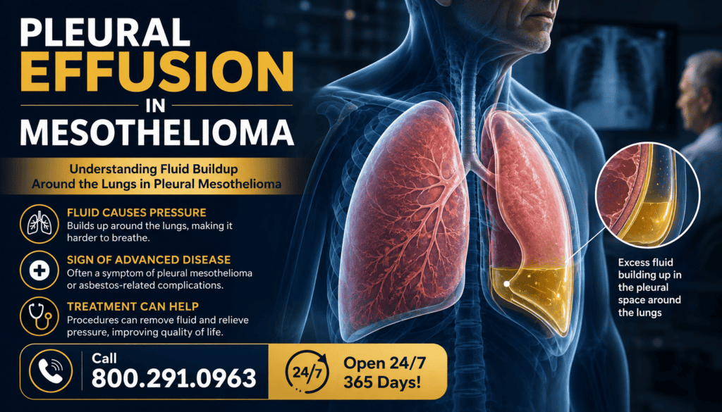
Pleural effusion means excess fluid has collected between the two thin pleural layers around a lung. In pleural mesothelioma, cancer involving this lining may interfere with normal fluid movement and contribute to a buildup that causes breathlessness, cough or chest discomfort. Fluid around the lung is a finding, not a diagnosis: infection, heart failure, blood clots and other conditions can also cause it. A clinician must determine the cause rather than assume every effusion in someone with mesothelioma is malignant.
Care may involve imaging, sampling or draining fluid, procedures to reduce recurrence, and treatment of the underlying illness. The best approach depends on symptoms, whether the lung re-expands, the fluid’s cause, the person’s goals and overall health. A sudden or severe breathing problem requires urgent medical evaluation.
🫁 What Pleural Effusion Means
The pleural space normally contains a small lubricating amount of fluid. An effusion is an abnormal excess. A large collection can limit lung expansion, but the volume of fluid does not always predict how breathless a person feels. Other conditions, including tumor affecting the lung or chest wall, can contribute to symptoms.
| Term | Meaning |
|---|---|
| Pleura | Thin membranes covering the lungs and lining the inside of the chest. |
| Pleural space | Potential space between the two pleural layers. |
| Pleural effusion | Extra fluid in the pleural space. |
| Malignant effusion | Fluid associated with cancer; cancer cells may be found in a sample. |
| Paramalignant effusion | Fluid in a person with cancer caused by an indirect mechanism rather than confirmed cancer cells in fluid. |
| Thoracentesis | Needle-and-catheter procedure used to collect or drain pleural fluid. |
| Pleurodesis | Procedure intended to seal pleural layers to reduce recurrent fluid. |
| Indwelling pleural catheter | Tunneled tube that permits intermittent drainage over time. |
Explore pleural mesothelioma and the broader mesothelioma symptoms guide.
🔬 Why Fluid Builds Up
Mesothelioma can disrupt the normal balance between fluid production and removal in the pleural space. Tumor-related inflammation, impaired lymphatic drainage and altered pleural surfaces can contribute. A patient may also have a separate cause of effusion; evaluation matters because treatments differ.
| Possible contributor | Why it matters |
|---|---|
| Pleural tumor involvement | Can disrupt normal fluid handling. |
| Inflammation | May increase fluid movement into the pleural space. |
| Impaired lymphatic drainage | Can reduce removal of fluid. |
| Pneumonia or pleural infection | Requires assessment and potentially antimicrobial treatment. |
| Heart failure | May cause fluid accumulation through a different mechanism. |
| Pulmonary embolism | Can cause breathlessness and sometimes an effusion; requires prompt assessment. |
| Treatment-related effects | Certain cancer therapies can contribute to fluid problems. |
| Low protein or other systemic illness | May contribute in selected patients. |
🩺 Symptoms & Warning Signs
Some small effusions cause no symptoms. Others can produce gradually worsening breathlessness, a dry cough or pressure and pain in the chest. Symptoms overlap with mesothelioma itself, infection, blood clots and other conditions. Contact the treating team about new or worsening symptoms rather than attributing them to fluid without evaluation.
| Symptom | What to do |
|---|---|
| Increasing shortness of breath | Contact the cancer team promptly; severe or sudden symptoms need emergency care. |
| Chest tightness or discomfort | Report location, onset and associated symptoms. |
| Dry or persistent cough | Discuss new changes and possible causes. |
| Difficulty lying flat | Tell the care team; note sleeping position and onset. |
| Fever or chills | Prompt medical review, especially with a drain. |
| Blue lips, fainting or severe breathing distress | Seek emergency medical care immediately. |
| New severe chest pain | Seek urgent evaluation; do not assume it is only an effusion. |
| Useful symptom record | Example |
|---|---|
| Onset | When breathing changed. |
| Activity | Breathlessness at rest versus walking. |
| Position | Whether lying down makes it worse. |
| Associated signs | Fever, pain, cough or swelling. |
| Prior drainage | Date, amount if provided by team, and whether it helped. |
Read more about shortness of breath, chest pain and chronic cough in mesothelioma.
🩻 Diagnosis & Fluid Testing
Clinicians use symptoms, examination and imaging to confirm fluid and look for its cause. Chest X-ray may identify an effusion; ultrasound can show fluid pockets and guide a safer procedure; CT may clarify pleural thickening, tumors or other findings. If sampling is indicated, pleural fluid can be examined for cancer cells, infection and chemical characteristics. A negative fluid cytology result does not rule out mesothelioma; tissue biopsy may be needed.
| Test | Purpose | Limitation |
|---|---|---|
| Chest X-ray | Shows fluid and other chest findings. | Does not establish why fluid formed. |
| Ultrasound | Locates fluid and guides drainage. | Does not independently prove malignancy. |
| CT scan | Examines pleura, lung and surrounding structures. | Imaging alone may not confirm diagnosis. |
| Thoracentesis | Obtains fluid for testing and may relieve symptoms. | Fluid testing may not identify mesothelioma. |
| Fluid cytology | Looks for malignant cells. | A negative sample cannot exclude cancer. |
| Pleural biopsy | Provides tissue for pathology when indicated. | Requires an individualized procedural decision. |
| Fluid test or assessment | Potential use |
|---|---|
| Cell count and differential | Assesses inflammatory pattern. |
| Protein and LDH | Helps characterize the fluid alongside blood tests. |
| Cytology | Searches for cancer cells. |
| Culture or other infection tests | Investigates infection when suspected. |
| Appearance and clinical context | Additional clues, not a diagnosis alone. |
| Pathology with appropriate markers | Can help distinguish mesothelioma from other malignancies. |
Related information: imaging tests for mesothelioma, mesothelioma biopsy and mesothelioma diagnosis.
💧 Thoracentesis & Drainage
Thoracentesis removes fluid through a needle or small catheter, usually with ultrasound guidance. It can provide a sample and sometimes improve breathing. The team may first review imaging, blood-thinning medications, bleeding risks and consent. The amount removed and monitoring depend on the patient and procedural circumstances. Fluid can return, so a single drainage is not necessarily a long-term solution.
| Stage | What patients can expect |
|---|---|
| Before | Review symptoms, medicines, imaging and procedure risks. |
| Positioning | Sit or lie in the position chosen by the clinical team. |
| Guidance | Ultrasound identifies an appropriate fluid pocket. |
| Local anesthetic | Numbs the insertion area. |
| Sampling/drainage | Fluid is removed for testing or symptom relief. |
| Afterward | Monitor breathing, pain and possible complications. |
| Follow-up | Review results and discuss recurrence prevention if needed. |
| Potential benefit | Important limitation |
|---|---|
| Relief of breathlessness | Not everyone improves; other causes may coexist. |
| Fluid for laboratory testing | Negative cytology does not exclude mesothelioma. |
| Assessment of lung re-expansion | May help select future procedures. |
| Short procedure in some settings | Bleeding, infection and pneumothorax are possible. |
| Can be repeated in selected cases | Repeated procedures may be burdensome if fluid rapidly recurs. |
For a procedure-specific explanation, see pleurocentesis (thoracentesis) for mesothelioma.
Need help now? Call 800.291.0963
Live chat is available 24/7, 365 days a year.
Submit Your Case
🔁 Recurrent Fluid & Long-Term Options
If fluid returns and causes symptoms, the care team may discuss repeat thoracentesis, an indwelling pleural catheter (IPC) or pleurodesis. Guidelines support either IPC or chemical pleurodesis for appropriate symptomatic patients whose lung expands. For nonexpandable lung, loculated effusion or unsuccessful pleurodesis, an IPC is generally favored over chemical pleurodesis. Decisions should reflect preferences, access to home support and treatment goals.
| Option | Potential role | Considerations |
|---|---|---|
| Observation | For selected people without symptoms. | Monitoring and reassessment are important. |
| Repeat thoracentesis | Intermittent relief or diagnostic sampling. | Fluid may recur; repeated visits and procedural risks. |
| Indwelling pleural catheter | Home or outpatient drainage of recurrent symptomatic fluid. | Requires supplies, training and infection monitoring. |
| Pleurodesis | Attempts to prevent fluid reaccumulation. | Works best when lung can expand adequately. |
| Treat underlying cause | Addresses cancer or another cause where possible. | Response and timing vary. |
| Supportive/palliative care | Relieves breathlessness and aligns care with goals. | Can accompany cancer-directed treatment. |
| Lung situation | Discussion point |
|---|---|
| Expandable lung | IPC and pleurodesis may both be reasonable. |
| Nonexpandable or trapped lung | IPC is often preferred to pleurodesis. |
| Separated fluid pockets | Imaging and specialist input guide feasibility. |
| Minimal symptoms | Intervention may not be needed immediately. |
| Limited caregiver support | Discuss realistic home-drain management. |
| Patient prioritizes fewer hospital visits | Consider durable options and trade-offs. |
Learn about pleurodesis and managing breathing problems and pleural effusion.
⚠️ Risks, Complications & Safety
All pleural procedures have potential harms. Thoracentesis can cause a pneumothorax (air around the lung), bleeding, infection or, rarely, problems related to lung re-expansion. Catheters can become blocked or infected. Pleurodesis can cause pain and fever and may not prevent recurrence. Ask the team which warning signs apply to the specific procedure and whom to call after hours.
| Concern | Warning sign or response |
|---|---|
| Pneumothorax | New or worsening breathlessness or chest pain after drainage: urgent assessment. |
| Bleeding | Persistent bleeding, dizziness or weakness: seek medical advice urgently. |
| Infection | Fever, redness, warmth, pus or increasing site pain: contact team promptly. |
| Catheter blockage | Unexpected loss of drainage with worsening symptoms: contact drainage team. |
| Fluid returns | Report recurrent breathlessness and discuss a longer-term plan. |
| Severe sudden symptoms | Emergency assessment rather than waiting for routine follow-up. |
| Home safety question | Why ask |
|---|---|
| Who drains the catheter? | Clarifies patient, caregiver or visiting nurse responsibilities. |
| How often should it be drained? | Use the team’s individualized schedule. |
| What is the dressing plan? | Reduces avoidable infection risk. |
| What symptoms require an immediate call? | Supports timely escalation. |
| Who supplies equipment? | Prevents interruption in care. |
| When is follow-up imaging needed? | Depends on symptoms and treatment. |
📋 Treatment Planning & Daily Care
Effusion care is part of a broader mesothelioma plan. The team may include an oncologist, pulmonologist, interventional pulmonologist, thoracic surgeon, nurses and palliative-care clinicians. Discuss the purpose of each procedure, expected symptom relief, how likely recurrence is, and whether home management fits the patient’s circumstances. Palliative care is symptom-focused support and can be provided alongside cancer treatment; it is not the same as hospice.
| Ask the care team | Reason |
|---|---|
| What is causing my fluid? | Different causes require different treatment. |
| Will drainage likely improve my breathing? | Other contributors may remain. |
| Did my lung fully re-expand? | Affects procedural options. |
| What if the fluid returns? | Establishes a clear next step. |
| Can I manage a catheter at home? | Requires training and support. |
| What symptoms need emergency help? | Makes the safety plan actionable. |
| How does this fit cancer treatment? | Coordinates symptom and disease management. |
| Can palliative care help now? | Adds symptom and family support. |
| Daily observation | What to record |
|---|---|
| Breathing | Changes at rest and with activity. |
| Pain | Location, severity and timing. |
| Temperature | Fever or chills when relevant. |
| Drain site | Redness, leakage or discomfort if a catheter is present. |
| Drainage | Only the amount/frequency directed by the clinical team. |
| Function | Sleep, walking, eating and daily activities. |
Related resources include mesothelioma treatment options, palliative care, symptom and fluid management and family caregiver support.
❓ Frequently Asked Questions
Is pleural effusion the same as mesothelioma?
No. Pleural effusion is extra fluid around a lung. Mesothelioma is a cancer of the mesothelial lining. Mesothelioma can cause an effusion, but fluid can have other causes.
Does fluid around the lung always mean cancer has spread?
No. Cancer can cause an effusion, but infection, heart failure, blood clots and other conditions may also be responsible. Testing and clinical evaluation establish the cause.
Can pleural effusion make breathing difficult?
Yes. Extra fluid can restrict lung expansion and contribute to shortness of breath, although other problems may contribute too.
How do doctors confirm the cause of the fluid?
They may use chest imaging, ultrasound-guided fluid sampling, laboratory studies and sometimes pleural tissue biopsy. A negative fluid cytology test does not rule out mesothelioma.
Will thoracentesis cure mesothelioma?
No. Thoracentesis drains fluid for diagnosis or symptom relief; it does not cure the underlying cancer.
What if the fluid keeps coming back?
The team may discuss an indwelling pleural catheter, pleurodesis or selected repeat drainage, based on symptoms and whether the lung expands.
Is a pleural catheter always better than pleurodesis?
No. Both may be reasonable for selected symptomatic patients with an expandable lung. A catheter is generally favored when the lung does not expand or pleurodesis has failed.
When should someone seek emergency care?
Sudden severe shortness of breath, blue lips, fainting or severe new chest pain warrant emergency evaluation. Contact the care team promptly for fever or concerning catheter-site changes.
🧾 Pleural Effusion in Mesothelioma Summary
| Key point | Practical takeaway |
|---|---|
| Definition | Extra fluid between the pleural layers around a lung. |
| Main symptoms | Breathlessness, cough and chest discomfort; some cases are asymptomatic. |
| Not automatically malignant | Cancer and noncancer causes must be distinguished. |
| Diagnosis | Imaging, possible fluid tests and sometimes tissue biopsy. |
| Initial drainage | Thoracentesis can sample fluid and relieve symptoms. |
| Recurring fluid | IPC or pleurodesis may be discussed for symptomatic patients. |
| Nonexpandable lung | An IPC is generally favored over chemical pleurodesis. |
| Treatment goals | Relieve symptoms, reduce burden and treat the cause when possible. |
| Emergency warning | Sudden severe breathing problems require urgent assessment. |
📚 Confirmed Medical & Clinical Guideline Sources
- National Cancer Institute — Cardiopulmonary Syndromes: Malignant Pleural Effusion
- National Cancer Institute — Mesothelioma Treatment
- National Cancer Institute — Mesothelioma Diagnosis and Prognosis
- American Thoracic Society / Society of Thoracic Surgeons / Society of Thoracic Radiology — Management of Malignant Pleural Effusions Clinical Practice Guideline (2018)
Source note: General malignant-effusion guidance is applied cautiously to mesothelioma; individual management depends on clinical findings. No disease-specific survival estimate is inferred from general malignant-effusion populations.
📨 Submit Your Case
Call 800.291.0963
Live chat is available 24/7, 365 days a year.
Submit Your Case
Having pleural effusion does not establish mesothelioma, asbestos exposure or eligibility for compensation. Diagnosis, exposure history, applicable deadlines and claim requirements must be evaluated individually. Compensation is not guaranteed.
⚕️ Legal & Medical Information Disclaimer
This article provides general information, not medical or legal advice. Seek emergency care for severe sudden breathing symptoms and consult qualified clinicians and legal professionals about individual circumstances.