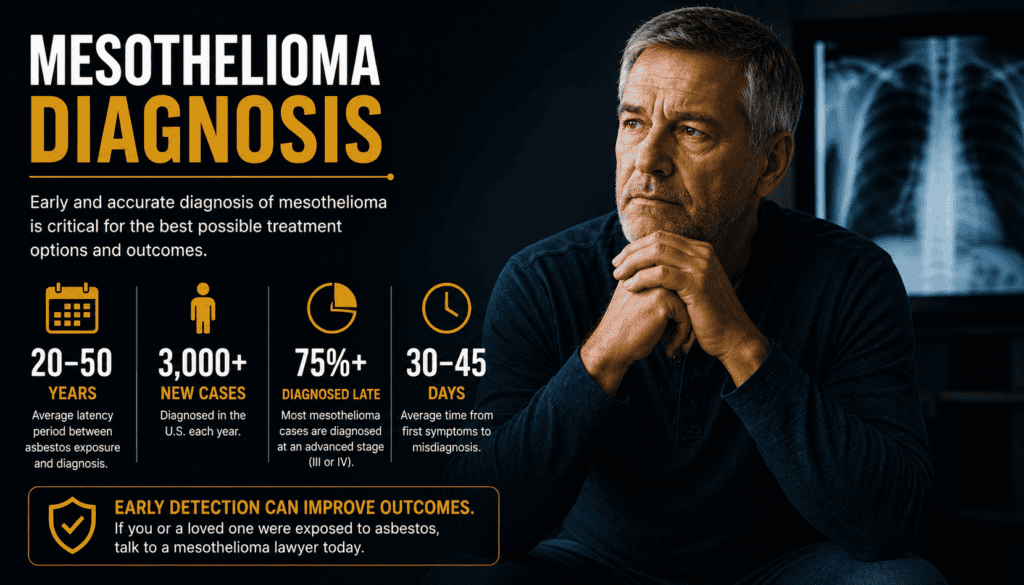
Mesothelioma Diagnosis
Tests, biopsy results, pathology and the steps from suspicion to a confirmed diagnosis
Mesothelioma diagnosis usually combines a medical history, imaging and laboratory examination of tissue. Because its symptoms and scan findings can resemble other illnesses, an abnormal image or a history of asbestos exposure does not prove that someone has mesothelioma. This guide explains what each test can and cannot tell you, what to ask about inconclusive results, and what happens after confirmation. Urgent symptoms: Seek emergency care for severe or sudden difficulty breathing, fainting, or severe chest pain. Contact your clinician promptly about new or rapidly worsening symptoms.
What Does a Mesothelioma Diagnosis Involve?
Diagnosing mesothelioma is a step-by-step process, not a single blood test or scan. Clinicians assess symptoms, exposure history and examination findings, use imaging to identify abnormalities, and obtain cells or tissue for a pathologist to examine. A suspicious scan is not the same as a confirmed diagnosis. Mesothelioma can resemble lung cancer or other conditions, making careful pathology particularly important. The sequence varies according to the suspected location, the person’s health and what earlier tests reveal. A patient with fluid around a lung may begin with chest imaging and fluid sampling; someone with abdominal swelling may need abdominal imaging and tissue sampling. Learn about the differences between pleural mesothelioma and peritoneal mesothelioma.
Preparing for a specialist appointment?
Learn how to collect medical records to support a mesothelioma diagnosis, including pathology reports, scans, biopsy records, and treatment notes.
| Step | Main question | Typical outcome |
|---|---|---|
| Clinical assessment | What symptoms and exposures warrant investigation? | A plan for imaging and appropriate referral. |
| Imaging | Where is the abnormality and how extensive might it be? | A target for sampling; imaging alone does not confirm the cancer. |
| Sampling | Can a suitable specimen be collected safely? | Fluid and/or tissue sent to pathology. |
| Pathology | What disease and cell type are present? | Diagnostic interpretation and possible additional stains. |
| Staging and planning | How far has confirmed disease spread? | Individualized discussion of treatment and supportive care. |
Symptoms, Asbestos History and First Appointment
Possible symptoms include persistent chest discomfort, breathlessness, cough, abdominal swelling, pain, fatigue or unintended weight loss. None is specific to mesothelioma; infections, heart disease, other cancers and benign causes can produce similar symptoms. Review the broader mesothelioma symptoms guide and tell the clinician when each change began. Discuss work, military, home-renovation and household exposure to asbestos if known. Exposure may have occurred decades before symptoms, and not recalling exposure does not exclude disease. Bring prior scans, pathology results, medication lists and a timeline of symptoms if available.
| What to bring | Why it helps |
|---|---|
| Symptom timeline | Shows onset, progression and effects on daily activities. |
| Employment and service history | Helps identify possible asbestos exposure without proving causation. |
| Prior imaging reports and images | Allows comparison with older findings. |
| Medication and allergy list | Supports safer test and procedure planning. |
| Questions and a support person | Helps clarify next steps and remember explanations. |
Imaging Tests: X-ray, CT, PET and MRI
A chest X-ray may show pleural fluid or thickening, but cannot establish mesothelioma. CT provides detailed cross-sectional images of the chest or abdomen and can help locate a biopsy target. PET/CT may help evaluate suspected spread in selected patients, while MRI can clarify invasion of nearby structures when clinically useful. The team selects tests based on the question being asked; not everyone needs every scan. Read more about imaging tests for mesothelioma. Tell your team about pregnancy possibility, kidney disease, contrast reactions and implanted devices when relevant. Imaging findings may raise or lower suspicion, but pathology is generally needed to distinguish mesothelioma from other conditions.
| Test | What it can show | Important limitation |
|---|---|---|
| Chest X-ray | Fluid or other chest abnormalities | Cannot identify mesothelioma definitively. |
| CT scan | Pleural or abdominal changes and possible biopsy targets | Some inflammatory changes can resemble cancer. |
| PET/CT | Metabolically active areas and possible spread | Inflammation can also show activity. |
| MRI | Detailed soft-tissue involvement in selected cases | Not routinely necessary for every patient. |
| Ultrasound | Fluid and guidance for some procedures | Limited for full staging on its own. |
Fluid Testing: Useful, but Not Always Conclusive
When fluid collects around a lung (pleural effusion) or in the abdomen (ascites), a clinician may remove a sample for laboratory analysis. Thoracentesis samples chest fluid; paracentesis samples abdominal fluid. Sampling may also relieve symptoms, depending on the circumstances. A cytologist examines cells in the fluid and may order additional testing. A negative fluid result does not rule out mesothelioma. Fluid may contain too few informative cells or fail to show the tissue architecture needed for a firm diagnosis. If suspicion remains, clinicians often pursue tissue biopsy. Likewise, a fluid result suggesting cancer may require further testing to establish exactly which cancer is present.
| Fluid finding | What it may mean | Reasonable follow-up |
|---|---|---|
| No malignant cells identified | No cancer cells seen in this sample | Discuss tissue biopsy if imaging and symptoms remain concerning. |
| Atypical or suspicious cells | Uncertain result | Ask whether more tissue or specialized tests are needed. |
| Malignant cells present | Cancer identified in fluid | Confirm origin and subtype with pathology as appropriate. |
| Fluid returns after drainage | Ongoing underlying cause | Discuss symptom control and definitive evaluation. |
Biopsy Methods and What to Expect
Biopsy removes tissue for microscopic examination. A CT-guided core needle biopsy may be possible when a safe target is accessible. Thoracoscopy uses a camera to examine the chest and sample suspicious pleura; laparoscopy serves a similar purpose in the abdomen. Some patients need a larger surgical sample when less invasive methods are inconclusive. The choice depends on location, bleeding risk, breathing capacity and the amount of tissue required. Before a procedure, ask whether you need to pause any medicines, whether sedation or anesthesia will be used, and when results are expected. Potential risks vary by procedure and can include bleeding, infection and, with chest sampling, pneumothorax (air around the lung). Your clinician should explain risks specific to you. See the dedicated mesothelioma biopsy guide.
| Procedure | Typical use | Key consideration |
|---|---|---|
| Image-guided core biopsy | Accessible chest or abdominal target | May yield limited tissue. |
| Thoracoscopy / VATS | Pleural inspection and multiple tissue samples | Requires procedural or surgical assessment. |
| Laparoscopy | Sampling suspicious abdominal lining | Often performed with anesthesia. |
| Open surgical biopsy | When other approaches cannot establish diagnosis | More invasive; selected circumstances only. |
Pathology, Cell Types and Diagnostic Uncertainty
A pathologist examines tissue structure and cell appearance, then may use immunohistochemistry and other tests to distinguish mesothelioma from metastatic cancer, lung cancer and reactive changes. Mesothelioma is commonly classified as epithelioid, sarcomatoid or biphasic. The cell type can influence treatment discussions, but a small specimen may not adequately represent the entire tumor. Ask for the final pathology report and whether a pathologist experienced with mesothelioma should review the specimen. If the report says “suspicious,” “atypical,” or “insufficient,” clarify whether the diagnosis is established or whether additional sampling is necessary. A second review may be particularly useful when the diagnosis is uncertain or treatment decisions are complex.
| Report term | Plain-language meaning |
|---|---|
| Final diagnosis | Pathologist’s interpretation of the tissue. |
| Epithelioid | One recognized mesothelioma cell type. |
| Sarcomatoid | A distinct mesothelioma cell type. |
| Biphasic | Both epithelioid and sarcomatoid components identified. |
| Immunohistochemistry | Marker tests that help distinguish similar-looking diseases. |
| Insufficient specimen | Not enough suitable material to answer the question reliably. |
After Diagnosis: Staging and Treatment Planning
Once mesothelioma is confirmed, the team determines its extent and assesses overall health. For diffuse pleural mesothelioma, TNM staging considers the primary tumor, lymph nodes and distant spread. Peritoneal mesothelioma is assessed differently; the pleural stage I–IV framework should not simply be copied to abdominal disease. See mesothelioma stages for a detailed explanation. Pathology, imaging, symptoms, functional status and patient preferences all inform care. A specialist team may discuss systemic treatment, selected procedures, clinical trials, symptom control and supportive services. Treatment is individualized; a stage number alone does not determine which option is appropriate. Review mesothelioma treatments and mesothelioma support resources.
| Question after diagnosis | Why it matters |
|---|---|
| Where did the cancer begin? | Distinguishes pleural from peritoneal and other rare locations. |
| What does the pathology confirm? | Establishes diagnosis and cell type. |
| Is additional staging needed? | Clarifies extent before treatment decisions. |
| Has a specialist reviewed the case? | Supports complex diagnostic and treatment decisions. |
| Which symptoms need relief now? | Symptom care can begin alongside further planning. |
Practical Checklist and Questions for Your Care Team
Request copies of radiology images, reports and pathology results; note who is responsible for ordering the next test and contacting you with results. If symptoms worsen while waiting, contact the care team rather than assuming the diagnostic process must finish first. New severe breathing difficulty, fainting or sudden severe chest pain warrants emergency evaluation. Costs and scheduling can be stressful. Ask about insurance authorization, travel support and a patient navigator. A trusted companion can take notes, and a written list of questions can make a short appointment more productive.
| Ask your clinician | What the answer clarifies |
|---|---|
| What is confirmed versus still suspected? | Prevents confusion between an abnormal scan and diagnosis. |
| Is tissue biopsy required? | Clarifies the next decisive test. |
| Is the sample sufficient for cell typing? | Identifies whether additional tissue may be needed. |
| Who will review my pathology? | Clarifies specialist involvement. |
| When and how will I receive results? | Sets a clear communication plan. |
| What symptoms require urgent attention? | Provides a safety plan while waiting. |
Frequently Asked Questions About Diagnosis
These answers provide general information; your care team can interpret your individual results.
Can a CT scan diagnose mesothelioma by itself?
No. CT can identify suspicious abnormalities and guide sampling, but imaging alone generally cannot confirm the diagnosis; tissue pathology is often necessary.
Does a negative pleural fluid test rule it out?
No. Fluid cytology can miss mesothelioma. If clinical suspicion remains, a clinician may recommend tissue biopsy.
Which biopsy is best?
There is no single best procedure for everyone. The choice depends on tumor location, accessibility, health and how much tissue is needed.
How long do biopsy results take?
Timing varies by laboratory, sample complexity and whether additional stains or expert review are needed. Ask your team for its expected turnaround and a contact plan.
Can mesothelioma be mistaken for lung cancer?
Yes. Some appearances overlap, so pathologists use tissue findings and specialized tests to distinguish them.
Do blood tests confirm mesothelioma?
No. Blood markers may contribute information in some settings but are not sufficient for diagnosis on their own.
Is a second pathology opinion reasonable?
It can be helpful when findings are uncertain or major treatment decisions depend on the diagnosis. Ask whether review by a mesothelioma-experienced pathologist is available.
Confirmed Medical References
Medical explanations in this guide are supported by these authoritative resources. Your own reports and clinician’s interpretation take precedence for individual care.
Get Help After a Mesothelioma Diagnosis
If you or a loved one has been diagnosed with mesothelioma and have questions about asbestos exposure, Call 800.291.0963. Live chat is available 24/7, 365 days a year. You can also Submit Your Case. Eligibility and compensation depend on individual circumstances; no outcome is guaranteed.
⚕️ Legal & Medical Information Disclaimer
This page provides general educational information, not medical or legal advice. A qualified healthcare professional should evaluate symptoms, diagnostic tests and treatment choices. Laws and filing deadlines vary; consult a qualified attorney for advice about a particular claim.